 Bright and early at 7:00 a.m., I arrive at the outpatient department of Jersey Shore University Medical Center, for my PET Scan. Standing there at the registration desk, with my large envelope of CT Scan films under my arm, I suddenly remember that I’ve forgotten to bring my prescription script. That’s kind of like showing up at the airport without your ticket. Early-morning brain freeze.
The round-trip back from Jersey Shore to our home is about 45 minutes, I think to myself. If I have to go back and fetch my script, it could set back the whole day’s schedule of PET Scans (knowing how expensive these scans are, that delay could be a really big deal).
Fortunately, after a few minutes of shuffling through various files, the clerk at the registration desk saves the day. It seems Dr. Lerner’s office faxed over a copy of the script a couple of weeks ago, as they booked the scan for me. Feeling much relieved, I move on to the radiology waiting area.
A technician comes and gets me, and ushers me down the hall and onto the trailer where the traveling PET Scan machine is housed (it will move on to another hospital tomorrow). The procedure is much the same as my last PET Scan, back on November 16th. A shot of radioactive glucose into my arm, a half-hour wait, then I take my place on the narrow, sliding table pointing into the aperture of the large, donut-shaped machine.
I lie on my back with my hands extended over my head – an uncomfortable position. With a low, whirring sound, I slide forward, feet first, into the scanner. I start out all the way in, with the top of the scanner just inches from my nose. Although I’m not especially claustrophobic, I find it easier to keep my eyes closed for this part.
After a few nearly-silent minutes of scanning, the machine slides me out a bit, head first, and pauses for another few minutes of scanning. After this interval, I slide out a little more, then I’m able – to my relief – to move my arms down and cross my hands on my chest. From this position, I can twist my neck and look over at a small, computer-type screen on the side of the scanner. Moments later, after the scan has re-commenced, I notice a message appearing in glowing letters, next to a flashing orange light: “Transmission source extended, remove non-essential personnel.” This evidently refers to whatever source of radiation makes the scanner work. I suppose I’m the only essential personnel here, right now (the technicians are back in their little room, safe behind a lead screen, no doubt). On the PET Scan trailer, "essential personnel" have the privilege of getting zapped with radiation. Fun.
The whole procedure – including registration and the wait time while the radioactive glucose makes its way through my body – takes about two hours, and is not unpleasant (except for the arms-over-the-head posture). I’m back home by mid-morning.
I spend some time today, off and on, thinking about something I just read on the narrative report from last week’s CT Scans – the ones I carried up to Jersey Shore today. Comparing this most recent scan to my prior scan of March 13th, the report reads: “Again noted is the spiculated retroperitoneal mass located at the level of each renal hilum. It is unchanged in size and continues to measure about 7cm in diameter. Additional small tiny retroperitoneal lymph nodes are also seen.”
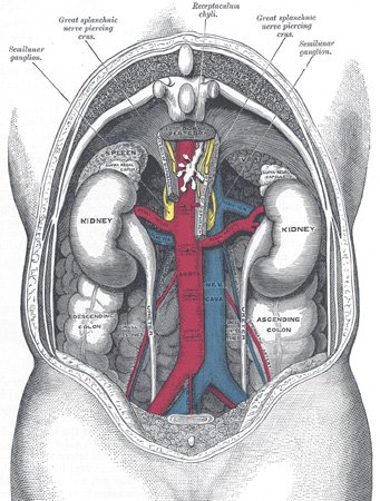 I suppose this must be the mass in my abdomen, that’s been the principal target of the chemotherapy treatments – but, not knowing the medical terminology, I can’t be sure. After some hunting around on the internet, I figure out that “spiculated” means “starburst-shaped.” “Retroperitoneal,” I know, means “towards the back of the abdomen.” “Renal” means having to do with the kidneys. “Hilum,” I discover, is the place where a duct or blood vessel enters an organ (in this case, the kidney).
Is this “the mass” we’ve been so concerned about? (It would seem so; I can’t think what else it could be.) If so, what are the implications of it being “unchanged in size,” after three additional chemo treatments? As of the last prior CT Scan, following chemo treatments 1, 2 and 3, the abdominal mass had shrunk by 50%. Is this most recent report saying it hasn’t shrunk any further, over the period of time covered by chemo treatments 4, 5 and 6? If so, is that significant? Or is this narrative report speaking of something else altogether? Too many questions, not enough answers.
Dr. Lerner warned us, earlier, that the mass won’t completely disappear, but will rather shrink down to the point where only scar tissue remains. Does the 7-cm width mean it’s now only scar tissue, or does it mean the chemo has somehow failed to shrink it further? The PET Scan results – with their ability to highlight “hot spots” of fast-growing tissue that are likely to be malignancies – may be just what we need, before anyone can say for sure.
 I don’t know whether or not I’ll get a phone call from Dr. Lerner in the next day or so, interpreting the CT scan results (although I suppose he would have called me by now, if he were going to). Maybe he’s waiting for the PET Scan results before phoning me – or maybe he’s just waiting for our appointment on the 31st, when he'll explain it all, face-to-face.
Until now, I’ve been content to just sit and wait for whatever news is going to come. Yet, having read the cryptic, dispassionate jottings of the radiologist, I’m eager, now, to have some explanation of what’s going on inside me – a description of not only what the CT Scan revealed, but what the full implications are.
It’s at times like this that we “health-care consumers” earn our more traditional label: patients.
1 comment:
Your writing makes us feel like we are right there with you but, of course, we do not ever pretend to know fully what you are experiencing. Thank you for being so open with us as you journey to wellness. We pray for answers for you that will lighten your burden. Charlene/Harvey
Post a Comment